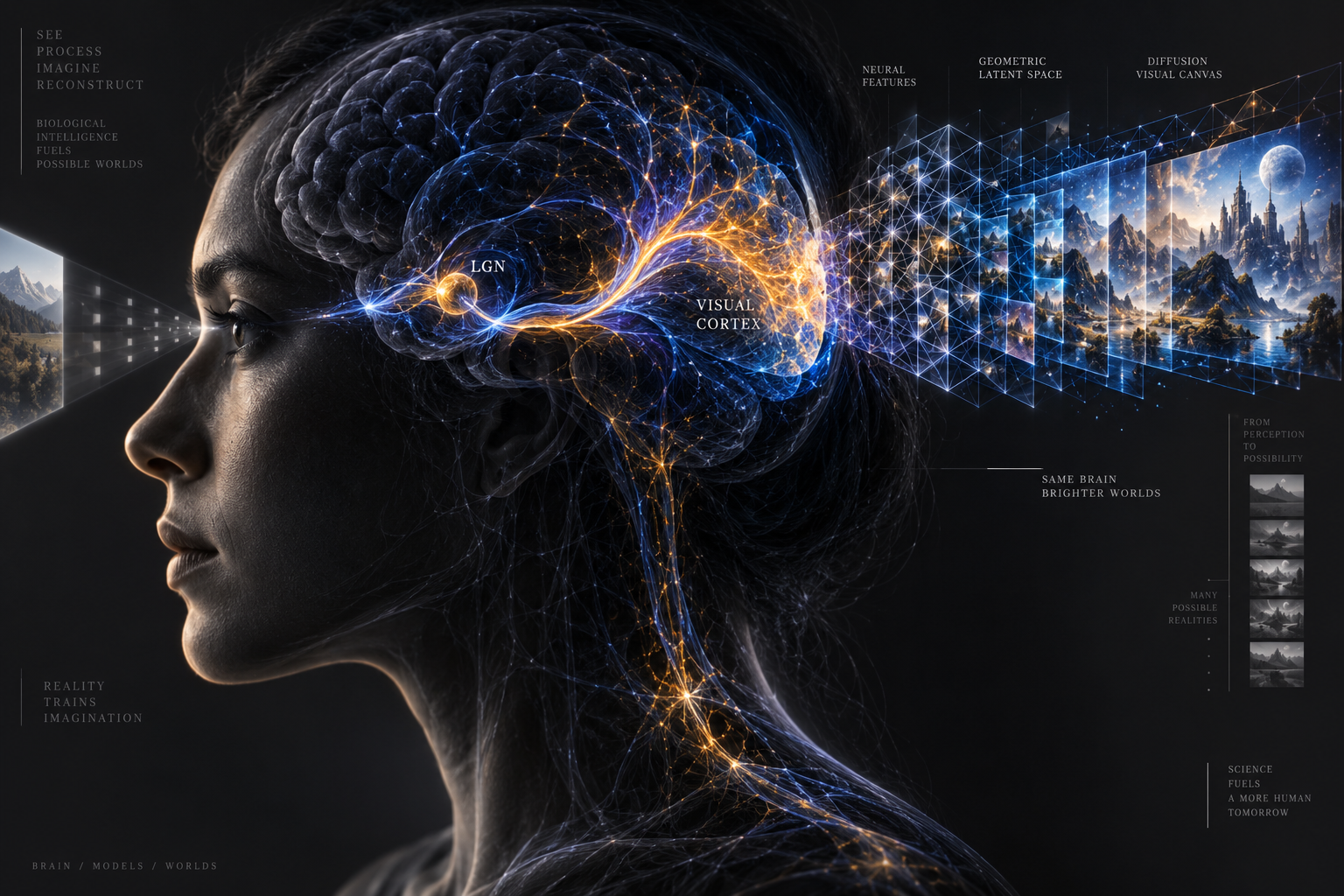
For more than two decades, cognitive neuroscience has chased a tantalizing ambition: decoding the subjective visual experience of the human mind directly from metabolic fluctuations in the brain. When someone gazes at a red sailboat cutting across choppy grey water, an intricate cascade of electrochemical impulses ripples through roughly one hundred million neurons in the primary visual cortex. Those neurons burn glucose and consume dissolved oxygen, triggering a localized compensatory rush of oxygenated hemoglobin through microcapillaries. Functional magnetic resonance imaging, or fMRI, registers this hemodynamic response as blood-oxygen-level-dependent (BOLD) contrast signals. Yet for most of the history of neuroimaging, translating those metabolic ripples back into the pictures that triggered them yielded little more than ghostly smears, ambiguous blobs, and crude spectral heatmaps.
That boundary shifted dramatically this week with the publication of new research from the Weizmann Institute of Science, covered in detail by MIT Technology Review on October 1, 2026. A research team led by computer scientist Michal Irani and doctoral researcher Roman Beliy unveiled Brain-IT, an architecture that bridges high-resolution neuroimaging and latent diffusion models. Where previous decoders produced uncanny approximations that merely guessed generic categories, Brain-IT reconstructs images that preserve both fine structural layout and precise semantic identity. If a volunteer inside the scanner looks at a specific black-and-white border collie resting on a wooden porch, the reconstructed image does not merely display a generic dog. It reproduces the exact posture, the orientation of the paws, the angle of the porch boards, and the contrast of light falling across the fur.
Predictably, the breakthrough reignited sensational headlines proclaiming that artificial intelligence has finally achieved telepathic mind-reading. Tabloid commentary warned of Orwellian interrogations where covert scanners pluck private memories and intimate thoughts from unsuspecting citizens. The truth of the matter is vastly more interesting than science fiction, grounded in rigorous bioengineering and strict physical limits. Understanding how Brain-IT works reveals why visual reconstruction represents a triumph of computational neuroscience, why modern diffusion models act as powerful visual prior engines rather than direct mind readers, and why the physical laws of biology keep your inner thoughts safe from surveillance.
🧠 The Sudden Leap from Fuzzy Blobs to Recognizable Images
To appreciate why Brain-IT marks such a pivotal milestone, one must examine the fundamental limitations that crippled earlier neural decoding attempts. In the late 2000s, pioneering researchers like Jack Gallant at the University of California, Berkeley, demonstrated that voxel activity in visual areas V1, V2, and V3 could be matched against a finite library of pre-recorded candidate videos. Gallant's system did not generate new images from scratch. Instead, it behaved like an elaborate search engine, selecting the best-matching clip from thousands of options already cataloged in a database. If the subject viewed something entirely novel, the system failed completely.
By the early 2020s, generative adversarial networks (GANs) and early diffusion pipelines allowed researchers to synthesize unconstrained images from brain signals. Yet these systems suffered from a persistent structural disconnect. A generative model trained on brain activity could reliably identify semantic categories (determining that the subject was looking at an aircraft, a mountain, or a human face), but it routinely placed the object in the wrong part of the frame, scrambled geometric proportions, or hallucinated wildly inaccurate backgrounds. If a subject looked at a red sports car on the left side of a highway, the reconstruction might render a yellow vintage truck in the center of an empty desert. The model understood the high-level concept of a motor vehicle, but the spatial coordinates of the visual field were completely lost in translation.
The core reason for this failure stems from the distinct functional organization of the human visual pathway. When photons hit the retina, signals travel through the lateral geniculate nucleus to the primary visual cortex (V1) at the back of the brain. Area V1 exhibits precise retinotopic mapping: adjacent points on the retina correspond to adjacent clusters of neurons in cortex. V1 acts as a low-level spatial grid, mapping edges, orientations, spatial frequencies, and contrasting boundaries. As visual signals travel forward along the ventral stream toward the inferior temporal cortex, the representation becomes increasingly abstract. Higher visual areas discard spatial pixel coordinates in favor of semantic categorization, recognizing that an animal is a feline regardless of whether it appears on the left, right, top, or bottom of the visual field.
Previous machine learning pipelines collapsed these distinct streams into a single homogenous embedding vector, forcing a generative network to guess both the layout and the identity from an entangled numerical stew. Brain-IT solved this problem by architecturally respecting the separation of labor in the human brain. By separating the reconstruction process into two dedicated, parallel branches—one for localized spatial geometry and one for categorical semantics—the system provides diffusion models with the exact dual guidance required to synthesize high-fidelity visual scenes.
🔬 How fMRI Actually Listens to the Visual Cortex
The primary window through which researchers observe this neural symphony is the functional magnetic resonance imaging scanner. A modern 3-Tesla or 7-Tesla research scanner is an engineering marvel, enclosing the volunteer in a massive superconducting solenoid chilled by liquid helium to near absolute zero. When the magnet engages, it aligns the magnetic moments of hydrogen protons in the water molecules that permeate human brain tissue. Radiofrequency excitation pulses tip these protons out of alignment; as they relax back to their baseline state, they emit tiny radio signals picked up by multi-channel receiver coils wrapped closely around the subject's head.
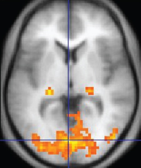
The critical biological hook is the difference between oxygen-rich and oxygen-poor blood. Deoxygenated hemoglobin contains unpaired electrons that make it paramagnetic, slightly distorting the surrounding magnetic field and accelerating the loss of signal coherence. Oxygenated hemoglobin, by contrast, is diamagnetic and leaves the local magnetic field undisturbed. When a cluster of neurons fires vigorously in response to a visual stimulus, local astrocytes and vascular smooth muscle dilate nearby arterioles, flooding the active tissue with fresh, oxygenated arterial blood in a phenomenon known as the neurovascular coupling response. This localized surge causes a measurable increase in magnetic resonance signal intensity: the BOLD signal.
However, the BOLD signal is not a direct electrical recording of neural spikes. It is a slow, indirect metabolic echo. While a neuron fires action potentials on a millisecond timescale, the hemodynamic response takes between four and six seconds to reach its peak after stimulus onset, lingering for several seconds before returning to baseline. Furthermore, standard fMRI resolution divides the cortex into three-dimensional volumetric pixels, or voxels, typically measuring between 1.5 and 2 millimeters on each side. A single voxel of that size contains between 50,000 and 100,000 individual neurons, along with millions of synapses, hundreds of capillaries, and extensive glial networks. Reading an fMRI scan is akin to attempting to decipher the dialogue of a theater performance by measuring the heat radiating from the roof of the auditorium five minutes after the scene concluded.
Extracting sharp visual details from this coarse, delayed hemodynamic wash requires immense mathematical sophistication. Previous decoders treated each voxel as an independent scalar variable or flattened the entire visual cortex into an undifferentiated vector. Brain-IT took a profoundly different route. Irani and Beliy organized voxels into functional clusters based on their retinotopic tuning and shared receptive fields across cortex. By modeling the interactions between these clusters, their Brain Interaction Transformer (BIT) captures how different regions of the visual hierarchy collaborate during active sight, transforming noisy hemodynamic readings into clean, localized feature maps.
🧩 Separating Spatial Layout from Semantic Meaning
The architectural heart of Brain-IT is its dual-stream interaction design. Rather than attempting to map brain voxels directly to final pixel coordinates, the Brain Interaction Transformer acts as an intermediate translator between neurobiology and computer vision primitives. It translates hemodynamic activations into two complementary feature sets that modern latent diffusion models natively understand.
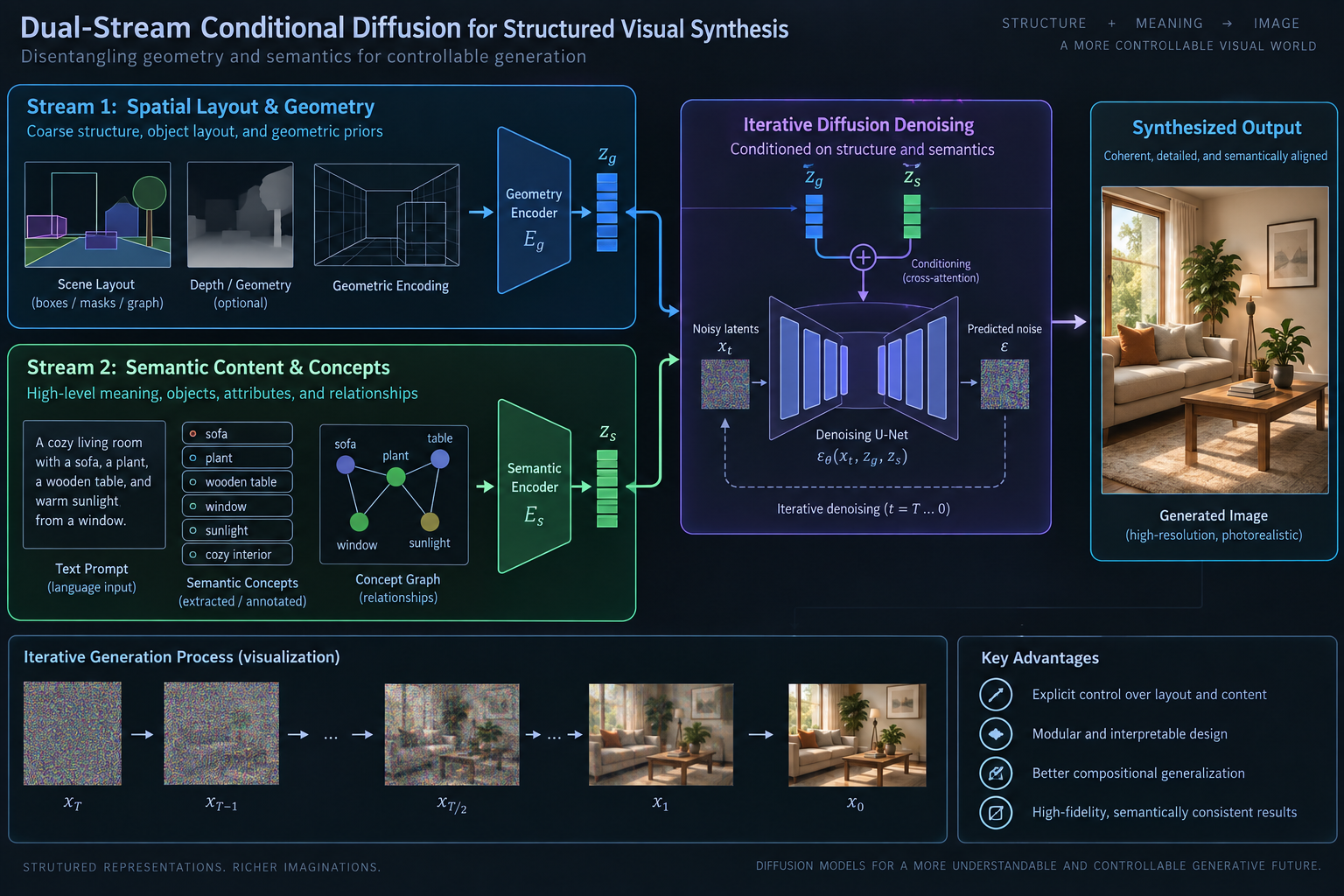
The first branch focuses exclusively on low-level structural layout. By reading voxels situated primarily in early visual areas V1, V2, and V3, the model extracts coarse spatial edge maps, luminance gradients, and patch-level color distributions. This structural guidance does not attempt to recognize what an object is; it merely records that a dark horizontal mass occupies the lower third of the canvas, a bright diagonal wedge cuts across the upper right, and a cluster of high-frequency edges is centered slightly left of middle. In computational terms, this provides the structural initialization for the reverse diffusion process, anchoring the composition to the exact physical layout observed by the retina.
The second branch focuses on high-level semantic tokens. This stream interrogates voxels located further along the ventral visual stream, including area V4, the lateral occipital complex, and the parahippocampal place area. These cortical regions respond selectively to semantic concepts such as faces, bodies, tools, natural landscapes, and architectural environments. The Brain Interaction Transformer maps these activations directly into the latent text-and-concept embedding space of a diffusion backbone, producing a rich categorical vector that specifies what entities exist within the scene.
When these two streams converge inside the diffusion architecture, the synthesis process is tightly constrained. The semantic stream prevents the model from generating visual gibberish, directing it toward authentic real-world objects and textures. Simultaneously, the structural stream prevents the model from exercising artistic license, forbidding it from moving objects, altering perspectives, or inventing arbitrary compositions. The result is a visual reconstruction that mirrors both the conceptual essence and the physical geometry of the original retinal image.
🎨 The Role of Diffusion Priors in Hallucinating Reality
To understand why Brain-IT produces such astonishingly crisp images, one must confront a crucial philosophical and technical distinction: the difference between signal extraction and generative synthesis. When viewing a side-by-side comparison of the original stimulus and the Brain-IT reconstruction, the generated image appears razor-sharp, complete with photographic textures such as water droplets on glass, individual dog whiskers, and the intricate weave of a cotton jacket. Does the fMRI scanner genuinely capture the microscopic texture of a dog's fur from blood flow inside the skull?
The rigorous answer is no. A 2-millimeter fMRI voxel is physically incapable of recording the high-frequency spatial frequencies required to render individual hairs or textile threads. Those photorealistic details are contributed not by the subject's brain, but by the generative prior embedded within the diffusion model.
A latent diffusion model, such as Stable Diffusion or its modern successors, has been trained on hundreds of millions of natural photographs. Through that training, it has internalized a vast statistical understanding of how the visual world is assembled: how light scatters through translucent leaves, how shadows pool beneath curved surfaces, and how skin texture varies across lighting angles. During the reconstruction process, the diffusion model begins with a canvas of pure Gaussian noise. Guided iteratively by the structural and semantic conditioning vectors produced by Brain-IT, the model systematically strips away noise, step by step, steering the emerging pixels toward a photograph that simultaneously satisfies the structural constraints from V1 and the semantic labels from higher visual areas.
In essence, the AI model acts as a skilled forensic sketch artist. The fMRI data provides the witness testimony: rough sketches of where shapes lie, broad notes on color palettes, and positive identifications of key subjects. The diffusion model provides the artist's professional mastery of anatomy, perspective, and lighting, filling in the microscopic details that the raw signal could never hope to carry. This synergy produces an image that is faithful in identity and geometry while appearing photorealistically complete.
Recognizing this mechanism is essential for dispelling the mythology of mind-reading. The algorithm is not projecting an internal mental movie reel onto an external monitor. It is synthesizing a new photograph that reconciles two distinct information sources: the coarse physiological footprints recorded by the magnet, and the statistical probabilities of natural imagery encoded within an artificial neural network.
⏱️ From Forty Hours to One: The Data Efficiency Breakthrough
Beyond the sheer visual quality of its outputs, Brain-IT achieved an equally consequential breakthrough in data efficiency. Historically, neural decoding pipelines were notorious for requiring absurd quantities of training data for every single participant. Because human brains exhibit substantial anatomical and functional idiosyncrasies—no two cerebral cortices fold in the exact same pattern, and visual field mappings vary across individuals—a decoder trained on Person A could not decipher brain scans from Person B.
To build an effective individual decoder, previous frameworks required volunteers to spend thirty, forty, or even sixty grueling hours lying motionless inside an fMRI bore across dozens of sessions, staring at tens of thousands of calibrated training images. This massive data burden restricted visual reconstruction research to small cohorts of healthy graduate students and dedicated research staff. It rendered clinical applications, where patients suffering from severe neurological trauma cannot endure hours of magnetic confinement, virtually impossible.
Brain-IT overcame this barrier by introducing functional voxel clustering that generalizes across distinct human subjects. Rather than treating an individual's brain as an isolated blank slate, the Weizmann team trained the core Brain Interaction Transformer on a collective foundation of publicly available neuroimaging datasets from multiple participants. The model learns universal functional relationships that govern how mammalian visual cortices integrate spatial and categorical information.
Because the structural transformer backbone is shared across individuals, adapting the system to a brand-new volunteer no longer requires forty hours of scanner time. The researchers demonstrated that with just a single one-hour calibration session—during which the participant views a modest set of standard images—Brain-IT aligns the new subject's idiosyncratic cortical coordinates into its shared functional space. The resulting reconstructions match or exceed the fidelity of legacy models that required weeks of exhaustive data collection. This reduction in calibration time from days to sixty minutes transforms neural decoding from an esoteric academic curiosity into a viable, deployable protocol for real-world clinical investigation.
🏥 Clinical Promise for Locked-In Patients and Neurorehabilitation
The true significance of high-speed, high-fidelity neural decoding lies not in novel consumer gadgets or speculative surveillance, but in restorative medicine. Across the globe, tens of thousands of individuals live with severe neurological impairments that strip away their ability to communicate with the outside world. Patients suffering from late-stage amyotrophic lateral sclerosis (ALS), extensive brainstem strokes, or catastrophic traumatic brain injuries can find themselves in a locked-in state: fully conscious, cognitively intact, and possessing vivid perceptual awareness, but entirely unable to move a single muscle, utter a sound, or track with their eyes.
For these individuals and their families, the loss of agency is devastating. While invasive brain-computer interfaces (such as intracortical microelectrode arrays pioneered by Blackrock Neurotech or Neuralink) have made inspiring strides in decoding motor intentions to control virtual cursors or robotic limbs, they carry the substantial risks of neurosurgery, tissue scarring, infection, and long-term electrode degradation. A non-invasive or minimally invasive decoding system capable of translating perceptual and mental states into intelligible visual and communicative tokens represents an extraordinary lifeline.
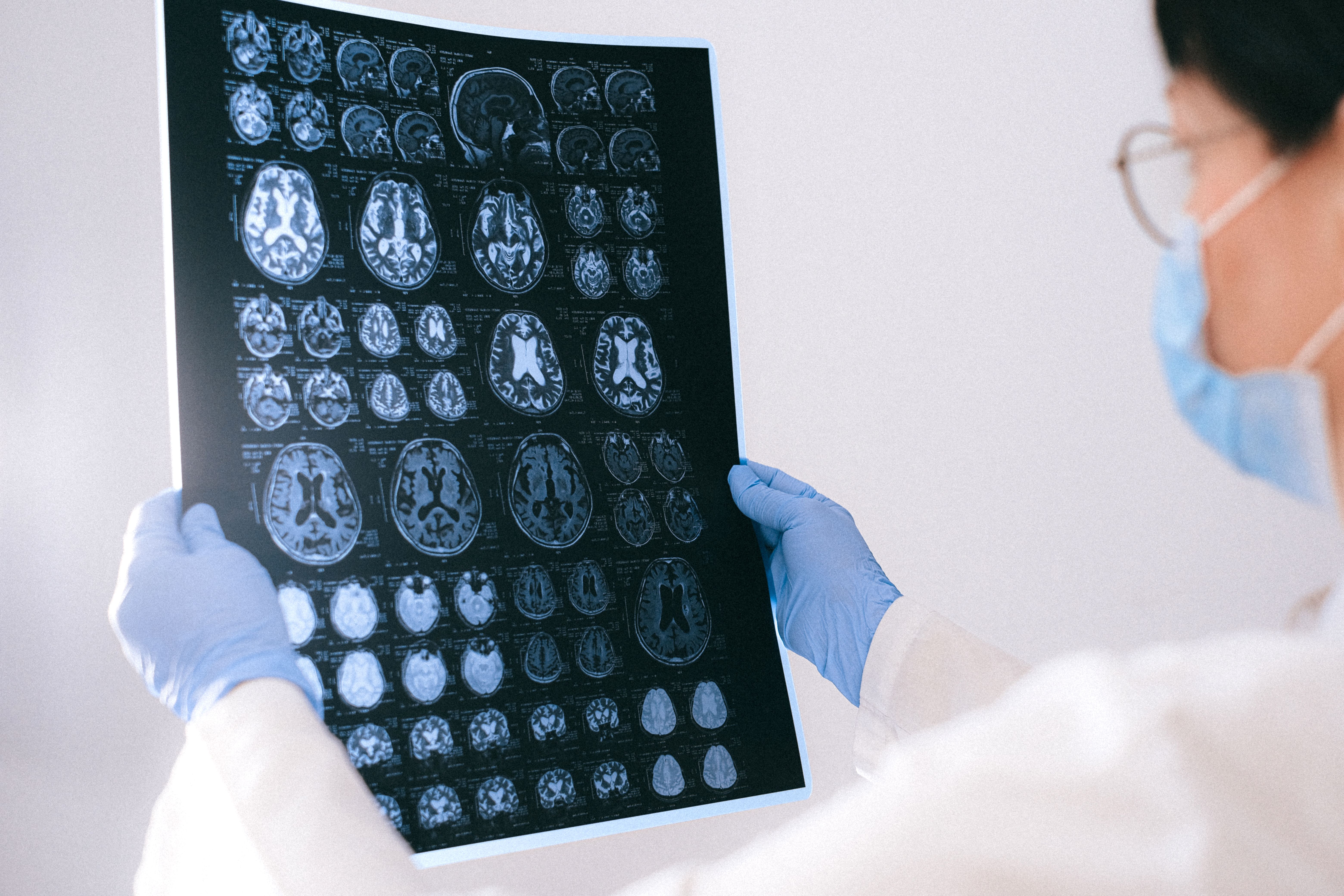
Visual decoders offer a direct diagnostic and communicative channel into the patient's internal experience. Clinicians can determine whether a minimally conscious patient is accurately perceiving their physical surroundings, recognizing the faces of loved ones, or processing educational and therapeutic materials. Furthermore, research teams are already extending the Brain-IT architecture from passive visual perception to active mental imagery and visual question answering (VQA). In early trials of "Brain-IT-VQA," researchers demonstrated that brain activations recorded while a subject mentally focuses on an object can provide answers to targeted questions about their visual environment.
Beyond communication for locked-in patients, high-fidelity neuroimaging decoders promise to revolutionize our understanding of sensory processing disorders, visual hallucinations in schizophrenia, and the neural substrates of sleep. For centuries, the visual content of human dreams remained completely inaccessible, locked inside the subjective recall of the waking dreamer. By deploying rapid, generalized decoders during rapid eye movement (REM) sleep, neuroscientists may soon capture direct visual reconstructions of oneiric imagery, shedding light on how the brain consolidates memories, rehearses survival behaviors, and processes emotional trauma.
🛡️ The Physics That Keep Your Private Thoughts Safe
Every time a study demonstrates enhanced neural decoding, it triggers an understandable wave of public anxiety regarding mental privacy. If an artificial intelligence can reconstruct the photograph a person is looking at from a brain scan, how long will it be before law enforcement, corporate employers, or hostile intelligence agencies scan our minds against our will?
The answer, rooted firmly in the physical laws of neuroimaging and the physiology of human cognition, is reassuring: involuntary mind-reading remains a technological impossibility under any plausible near-future framework. The barriers protecting cognitive privacy are not merely legal or regulatory; they are structural, physical, and biological.
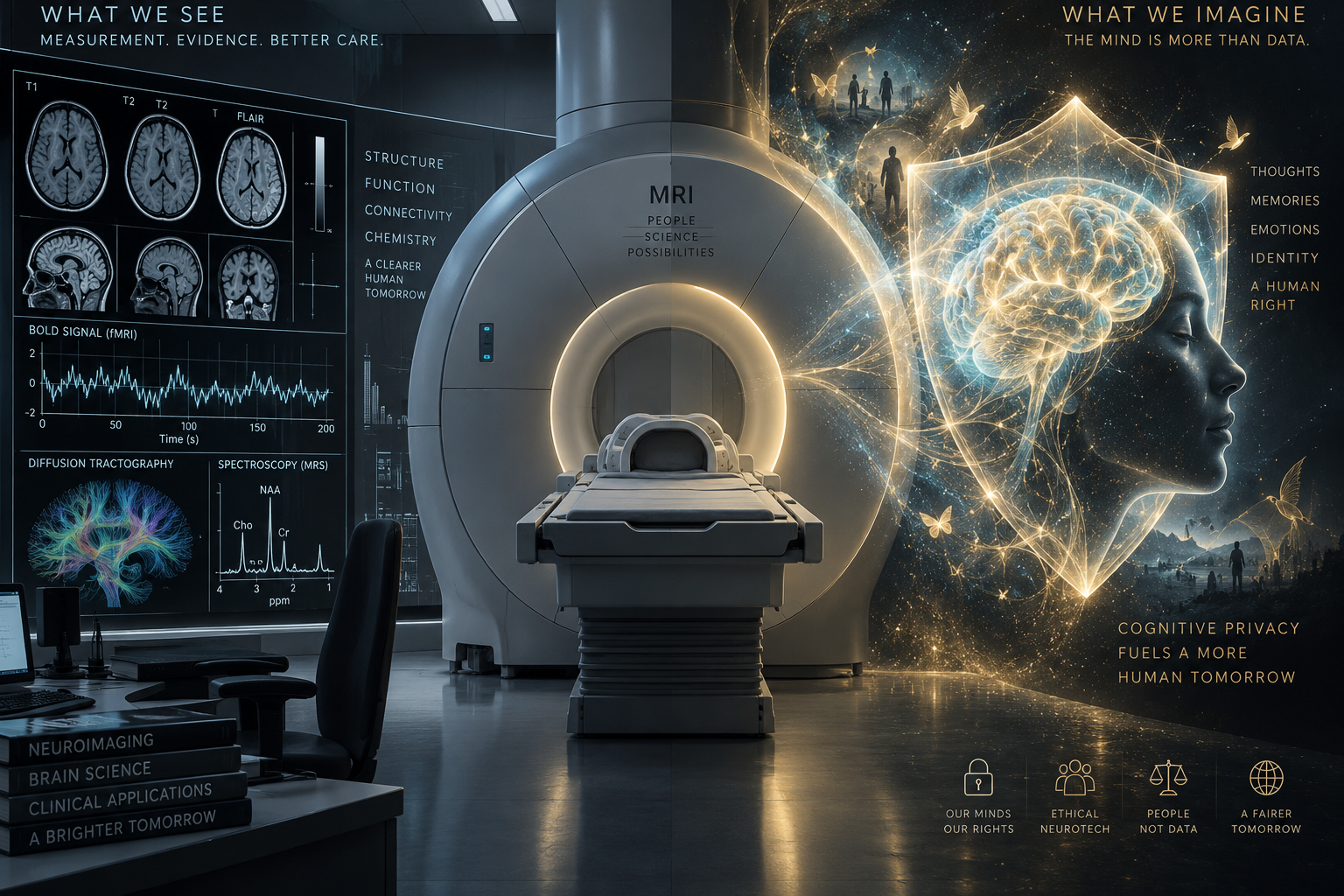
The first insurmountable barrier is hardware geometry. An fMRI scanner cannot be miniaturized into a discreet overhead camera, a ceiling tile, or a pair of lightweight sunglasses. Generating the stable 3-Tesla or 7-Tesla magnetic fields required to detect tiny hemodynamic shifts demands a massive cylindrical bore weighing between four and twelve metric tons, wrapped in vacuum-insulated cryostats containing thousands of liters of liquid helium. The subject must be slid into the physical center of this magnetic tube, surrounded by high-powered gradient coils that switch magnetic fields with ear-splitting acoustic thumps reaching 110 decibels. You cannot scan someone's brain without their absolute, physical immersion in a specialized multi-million-dollar medical facility.
The second barrier is motion sensitivity. High-resolution fMRI scans acquire data slice by slice over several minutes. If a participant moves their head by as little as two or three millimeters—the breadth of a single voxel—the spatial alignment collapses, transforming the data into uninterpretable spatial blur. Even natural physiological movements, such as heavy breathing, swallowing, or muscular clenching of the jaw, introduce severe motion artifacts that ruin the decoding pipeline. A subject who does not wish to be scanned can effortlessly neutralize the entire process simply by swallowing repeatedly, clenching their teeth, or shifting their neck.
The third and most profound defense is cognitive sovereignty. The human visual cortex responds dynamically to attention, focus, and internal cognitive tasks. If a subject lying inside the scanner is instructed to look at an image on a projection screen, but deliberately chooses to close their eyes, perform complex mental arithmetic, silently recite poetry, or daydream about their childhood home, the retinotopic activation in areas V1 through V4 instantly dissolves. The primary visual cortex requires active retinal engagement and focused visual attention to produce coherent spatial feature maps. You cannot steal images from a mind that refuses to look.
Finally, there is the fundamental biological distinction between passive visual perception and abstract inner thought. The visual cortex processes sensory inputs that are spatially organized across the cortical surface. In contrast, complex human beliefs, autobiographical memories, abstract moral reasoning, and covert verbal reflections are not stored as tidy two-dimensional pictures pinned to a cortical map. They emerge from non-localized, highly dynamic, multi-scale network interactions distributed across billions of neurons in prefrontal, temporal, and parietal networks. Decoding a picture of a coffee mug from the visual cortex is as different from reading someone's political convictions or financial secrets as measuring the temperature of a computer CPU is from decrypting its encrypted database.
⚖️ Neurorights and the Governance of Cognitive Privacy
Yet the impossibility of clandestine, sci-fi mind-reading does not excuse technological complacency. While involuntary surveillance remains a fantasy, the rapid consumerization of consumer-grade neurotechnology—such as non-invasive electroencephalography (EEG) headbands, functional near-infrared spectroscopy (fNIRS) caps, and earbuds embedded with neural electrodes—demands robust, proactive governance.
Over the past three years, international bodies including UNESCO and the OECD have sounded urgent warnings regarding the commercial collection of brain metrics. When consumer headsets track attention spans, cognitive fatigue, emotional arousal, or subconscious visual reactions to corporate advertising, that data enters commercial clouds largely unprotected by traditional healthcare privacy statutes like HIPAA. In several jurisdictions, neurodata collected outside a hospital or clinical trial is treated as ordinary consumer telemetry, vulnerable to being harvested, profiled, and monetized by digital advertising conglomerates.
A pioneering global movement spearheaded by neurobiologist Rafael Yuste and the NeuroRights Foundation has argued that existing human rights frameworks are ill-equipped to address technologies that directly interface with neural activity. They propose five fundamental "Neurorights" that must be codified into international and national law:
- The Right to Cognitive Liberty: The fundamental freedom of an individual to control their own mental processes, choose their state of mind, and refuse any unwanted neurotechnological intervention.
- The Right to Mental Privacy: The absolute protection of neural data, guaranteeing that the recorded electrical or metabolic activity of a human brain cannot be accessed, stored, or analyzed without explicit, informed, revocable consent.
- The Right to Psychological Continuity: Protection against technological manipulations that alter an individual's personal identity, personality, or core sense of self.
- The Right to Fair Access to Cognitive Augmentation: Ensuring that human enhancement technologies do not exacerbate socioeconomic inequality or create stratified castes of biologically augmented elites.
- Protection Against Algorithmic Bias in Neurotechnology: Mandating that neural decoders and neurotechnological models are audited for demographic fairness and trained on representative, unbiased cohorts.
Chile made history as the first nation to amend its constitution to explicitly safeguard brain data and cognitive integrity as fundamental human rights, followed by emerging legislative initiatives across Brazil, Spain, and the European Union. As machine learning models like Brain-IT demonstrate how effortlessly artificial intelligence can bridge biological signals and synthetic visual media, the global community must ensure that the legal boundaries around human consciousness remain inviolable.
The true triumph of Brain-IT is not that it breaches the sanctity of the human mind, but that it demonstrates how computational intelligence can decipher the elegant visual grammar of biology. By respecting the natural architectural division between spatial geometry and semantic meaning, researchers have built a powerful tool for neuroscience and clinical rehabilitation. The technology gives voice to those who cannot speak, maps the intricate circuitry of sight, and illuminates the deep computational principles that unite human and machine perception. Through it all, the physical reality of the human brain remains unchanged: open to healing, capable of communication, and fiercely, beautifully sovereign.
Sources: Weizmann Institute of Science Department of Computer Science and Applied Mathematics; International Conference on Learning Representations (ICLR 2026); MIT Technology Review; Nature Neuroscience; UNESCO Neurotechnology Governance Guidelines.